INDUSTRY NEWS
How Advanced Imaging is Elevating Medicine For Exotic Species typographic title
By Copper Aitken-Palmer, DVM, MS, PhD, Diplomate ACZM
T

hose of us that care for unique species rely on every diagnostic tool in existence, and even then, we still have to get creative for our patients. For many of our species, we do not have a known normal for comparison or reported reference ranges to rely on. Even as a Board-Certified Specialist in Zoological MedicineTM, this can feel like a limitation to the care I can provide to my patients. The species we care for vary widely. In a single day, a veterinarian caring for non-domestic species might examine a rabbit, a bearded dragon, a blue and gold macaw, and an echidna. Each has vastly different physiology, anatomy and disease prediction.

Powerful Diagnostic Tool
In recent years, there has been one diagnostic advancement that provides unparalleled consistency across species. This single diagnostic has changed how Zoological Medicine is practiced and has empowered us to provide better care. This diagnostic is Computed Tomography (CT). As advanced imaging with CT has become more readily available for veterinarians caring for zoo, exotic and wildlife species, we are learning its true power. It has changed the level of medicine we can provide, resulting in rapid advancements in our field.

Detailed Anatomy

Rarely do we have anatomy books to reference our species, especially when it comes to comparing specific organ anatomy across similar but different species. As an example, there are no reference books comparing anatomy among insectivores. Species that may seem similar, such as Giant anteaters, tamandua, or Asian and African pangolins, can have different adaptive anatomy. Hedgehogs are insectivores too, but there are undescribed differences between African hedgehogs and European hedgehogs.

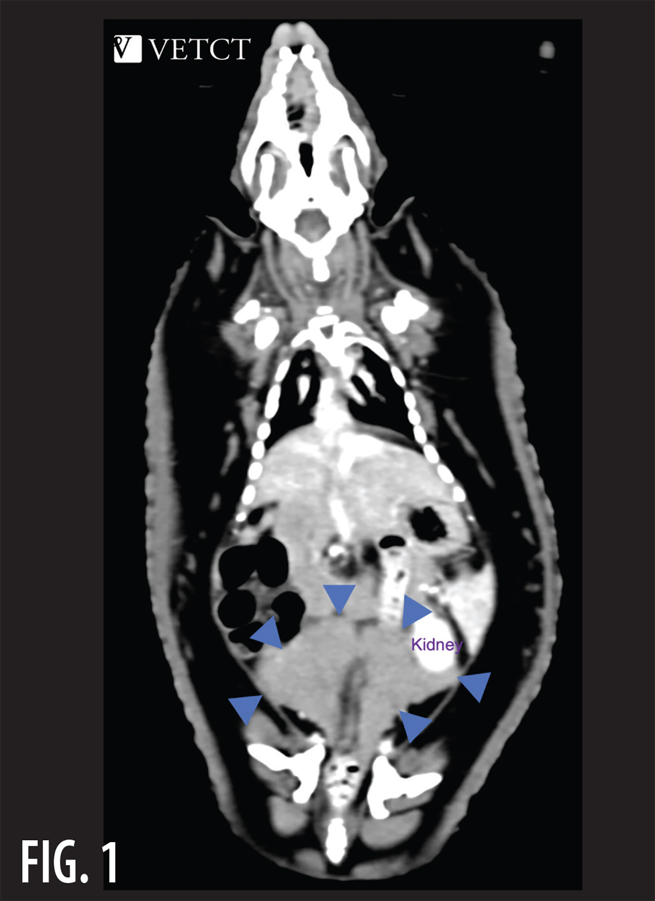
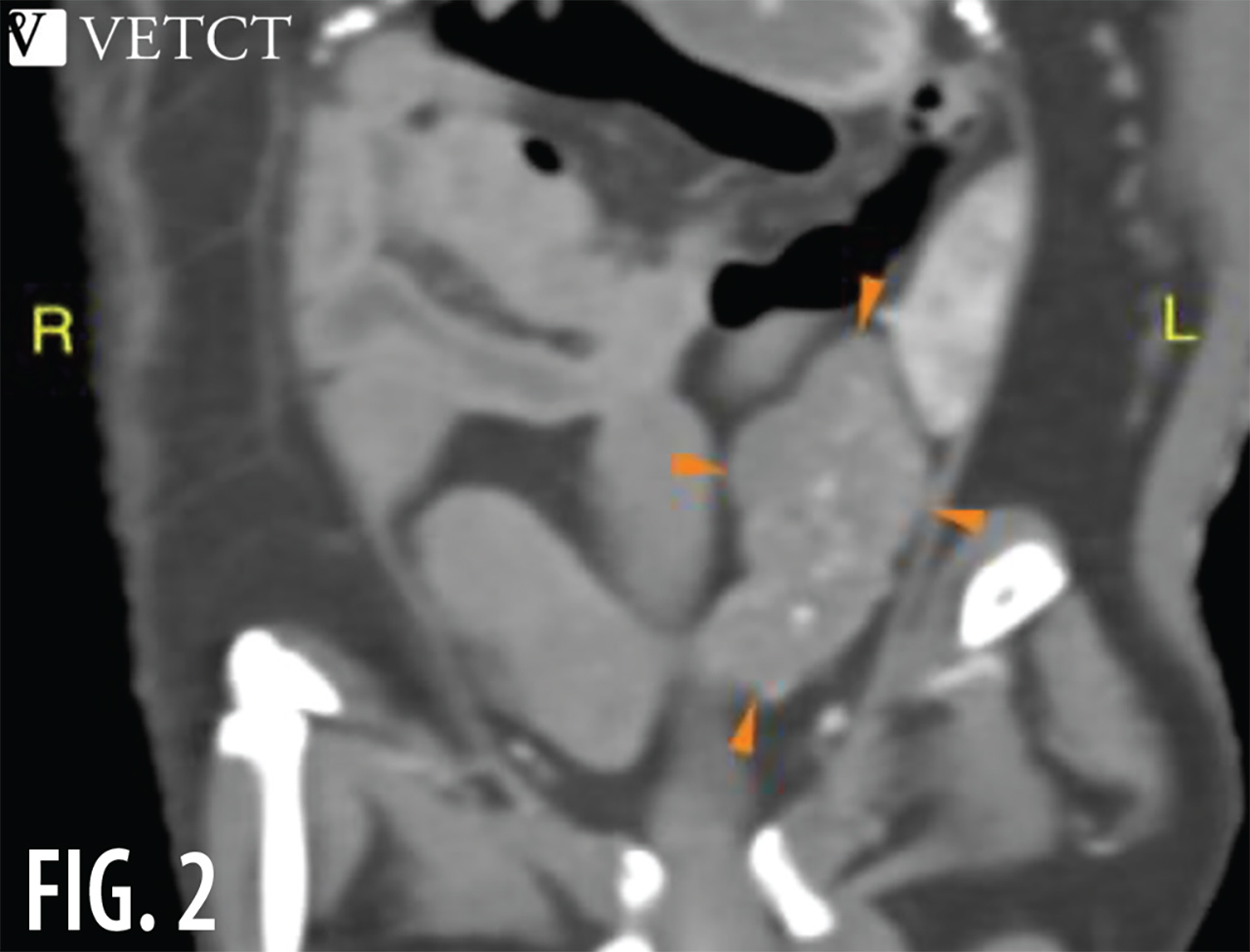
Computed Tomography with contrast enhancement has opened a new chapter to understand the anatomy of these species antemortem.1 Searchable databases are currently under development. Once we know the normal anatomy, it helps us better recognize abnormal disease changes when they occur.2,3

This is demonstrated in Fig. 1 of a suspected normal hedgehog and Fig. 2 of a suspected abnormal hedgehog CT with contrast enhancement. In the suspected normal CT image, the dorsomedial and ventrolateral lobes of the vesicular glands (blue arrows) are symmetrical with no mineralization. The hedgehog that is suspected to be abnormal (orange arrows) has asymmetry of the vesicular glands and visible mineralization of the left gland. The shape is different between the two images because of the overlapping dorsomedial and ventrolateral lobes of the vesicular glands in the normal animal. Without CT, we would not have been able to make an antemortem diagnosis for this hedgehog.

Consistent Across Species

There are no known reference intervals for normal body measurements for many species. Among reptiles, intraocular pressure (IOP) ranges are known for only a few species. It is hard to determine if these known values can be extrapolated from a green iguana to a savannah monitor—a distant taxonomic relative. These two species are in different families, as comparable as a cat and a dog. With CT, we do not need to know normal intraocular pressure. A working diagnosis of glaucoma can be made by differentiating acquired buphthalmia and exophthalmia with an eye that appears large. CT can reveal retrobulbar causes of exophthalmia, which if not present, indicates glaucoma causing buphthalmia is most likely. With CT, this is a diagnosis that can be made without normal IOP references.
Swift Diagnosis
Screenshot of a medical CT scan displaying a suspected normal hedgehog anatomy structure area
Screenshot of a medical CT can displaying a suspected abnormal hedgehog anatomy structure area with contrast enhancement
When used with contrast enhancement, CT also allows for swift diagnosis of complex medical conditions. A tiger must be anesthetized for any comprehensive exam. Obtaining radiographs of a tiger can be physically difficult, as the large animal is manipulated in the precise positions for each radiograph. This can add an hour to anesthesia time while working to get all the necessary views completed. Longer procedures while under anesthesia increase risk of hyperkalemia in tigers, making short procedures preferable.4 With CT, the tiger only needs to be positioned in sternal, and in less than 15 minutes detailed imaging of the entire body is obtained. With those images we can scan for abnormalities head to tail, diagnosing dental disease, rhinitis, neck arthritis, lumbar arthritis and cystic endometrial hyperplasia. No other single diagnostic provides such comprehensive diagnostic power with such ease and speed of acquisition.
Identifying New Conditions

Among our special species, previously unreported conditions are now identified with the help of contrast-enhanced CT. Understanding these conditions allows us to identify unexpected etiology and provide better care to our patients. Working with radiologists skilled in reviewing CT for exotic species has led to the rapid diagnosis and surgical correction of gastric dilatation and volvulus in rabbit, a previously unreported condition for the species.5 Among non-domestic species, this condition is often considered fatal because we were previously unable to diagnose and surgically correct it in time.6,7,8

It can be a challenge to coordinate a CT and image review rapidly enough for successful surgical correction for a condition like GDV. However, the increased availability of CT combined with the use of teleradiology allows us to rapidly access radiologists with exotics experience so that we can receive results directly no matter what time zone or time of day/night.

Extraordinary Detail
Rabbit and rodent skulls have many overlapping structures all within a small space. Dental disease and malocclusion can affect other organs such as eyes, nasolacrimal ducts, sinuses or ear canals, all in proximity. Multi-view radiographs can help identify these changes, but CT gives far superior detail. Simply, we miss abnormalities with radiographs that are easily seen with CT. Pulpitis, lens luxation, reserve crown infection, osteomyelitis, nasal mineralization and nasolacrimal narrowing can all be detailed with a single CT. And then response to therapy can be monitored with serial CT over time.
Genuine Breakthrough
CT is proving to be a genuine breakthrough diagnostic for those of us providing care for zoo, exotics and wild species. It is enabling us to identify and treat a range of abnormalities in a wide variety of species with a certainty we were previously lacking. However, while it is proving to be an essential diagnostic tool for certain species and conditions, it is not available to all. Cost, expertise and location can all be prohibitive to using this imaging modality. Improved access to CT machines, training in image acquisition and expert interpretation through teleradiology will increase access to CT imaging. This, in turn, will allow us to provide bettercare for more of our unique species.
References
  1. Verhulst, C., Aitken‐Palmer, C., & Holliday, C. (2019). New Imaging Approaches Enable Visualization of 3D Musculoskeletal Anatomy of African White‐bellied Pangolin. The FASEB Journal, 33(S1), 613-8.
  2. Zoo and Aquarium Radiology Database (ZARD), an Innovative Imaging Database Designed to Support Wildlife and Zoo Professions, proceedings IAAAM 2022, Eric T. Hostnik1,2*; Michael J. Adkesson1; Matt Kinney3
  3. “Free online collection of veterinary diagnostic imaging cases and hematology images of zoo animals.” www.imaios.com/en/zoo-paedia
  4. McEntire, M. S., Ramsay, E. C., Price, J., & Cushing, A. C. (2020). The effects of procedure duration and atipamezole administration on hyperkalemia in tigers (Panthera tigris) and lions (Panthera leo) anesthetized with -2 agonists. Journal of Zoo and Wildlife Medicine, 51(3), 490-496.
  5. Imrie, P. (2022). Vets Need to Be Aware of Rabbit GDV. Vet Times. www.vettimes.co.uk/news/vets-need-to-be-aware-of-rabbit-gdv-danger/
  6. Hinton, J. D., Aitken-Palmer, C., Joyner, P. H., Ware, L., & Walsh, T. F. (2016). Fatal gastric dilation in two adult black-footed ferrets (Mustela nigripes). Journal of Zoo and Wildlife Medicine, 47(1), 367-369.
  7. DiGeronimo, P. M., Enright, C., Ziemssen, E., & Keller, D. (2023). Fatal gastric dilatation and volvulus in three captive juvenile Linnaeus’s two-toed sloths (Choloepus didactylus). Journal of Zoo and Wildlife Medicine, 54(1), 211-218.
  8. Hinton, J. D., Padilla, L. R., Joyner, P. H., Schnellbacher, R., Walsh, T. F., & Aitken-Palmer, C. (2017). Gastric dilatation volvulus in adult maned wolves (Chrysocyon brachyurus). Journal of Zoo and Wildlife Medicine, 48(2), 476-483.
Portrait headshot photograph of Dr. Aitken-Palmer grinning in a dark navy blue dress shirt
Dr. Aitken-Palmer has been a veterinarian for 20 years. As a zoo, exotic, and wild animal specialist, she is passionate about improved veterinary care for unique species. She is known for her scientific contributions regarding male giant panda reproductive physiology and contributing to the growth of the giant panda population through improved artificial insemination. Dr. Aitken-Palmer speaks with veterinarians all over the world daily to provide specialized veterinary support as a telespecialist with VetCT. She works closely but remotely with a team of radiologists and other veterinary specialists at VetCT to empower first-line veterinarians that care for all species, from rabbits in the UK and tegu in the USA to hyena in the Middle-East.